Foam drainage experiments

Stephan Koehler & Eric Weeks -- <koehler(at)seas.harvard.edu> & <weeks(at)physics.emory.edu>

Lab Home -- People -- Experimental facilities -- Publications -- Experimental pictures -- Links

soap foam We wanted to see how liquid drains through foams. The picture at left is a simple soap foam, generated by producing bubbles in the liquid at the bottom. The liquid has some fluorescent dye in it, that's why it's yellow. You can see how all the bubbles are similar sizes, and thus pack in geometrically pleasing ways.

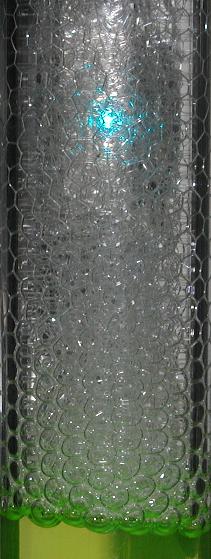
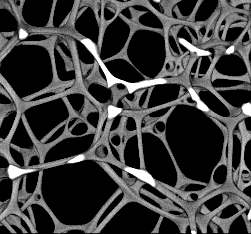
We trickled in some additional soapy water at the top of the experiment, and watch this water flow through the soap film by looking at an individual channel (where three bubbles meet) within the soap film with a confocal microscope. In fact, we put particles in the added water, so we can see them moving through the channel. From this we can extract velocity profiles within the channel, thus teaching us how water flows through the foam. The blue spot in the picture at left shows light from the confocal microscope laser.

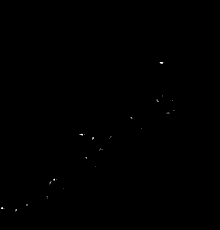
The key question is, do the soap bubble walls act like rigid walls, or like slippery walls? If they're rigid, then the water flows more slowly through the channels, and the flow will be fastest at the center of the channel, and nearly zero at the bubbles. If they're slippery, then the flow will be the same across the channel, and thus still quite rapid near the channel walls (the bubble surfaces).

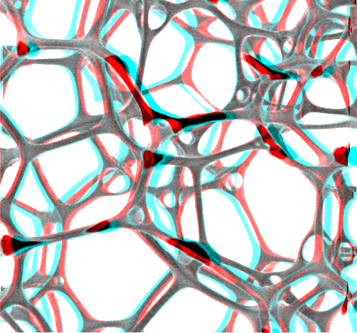
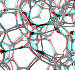
The picture to the right shows an image of the foam, displayed on the monitor, while we're taking data.
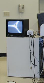
To do this experiment, we flipped the microscope on its back side, as shown in the picture below. Bud Puckett, the physics department machinist, built a nice platform for the microscope, and some Plexiglas holders to help hold the foam tube to the microscope stage. The yellow liquid is the soap solution, the foam is in the tube above the solution, with a hose running off the top to allow the excess foam to drain away (since we continually produce foam from below). You can see a small hose coming into the side of the foam tube, near the microscope, where we inject the liquid.


Click here to see an animated GIF movie of particles flowing through a foam in our experiment.

You can see that the flow is much faster near the centers of the channels, indicating that the bubble walls are behaving rigidly in this experiment.

This foam was made using BSA, a common protein, rather than some other surfactant (soap) molecule. (BSA is "bovine serum albumin".) Gravity points downwards (that is, the flow you'll see is caused by gravity), and the width of the picture is 0.5 mm. The movie represents 3 seconds of data. The particles are 1 micron in diameter.


Two movies of flow in a channel. 2.5 s of data, the surfactant is Tween 20. On my web browser, these two movies don't play back at a steady rate (due to limitations of the animated GIF format); the flow is constant for this data set, despite the movie playback.

Another movie, showing flow from one channel to another. We think the white blob is due to a stagnation point in the flow, where a lot of fluorescent particles are collecting. This movie is 3 s of data.


Three-dimensional movies

This is a movie showing the structure of an emulsion sample, rotated in 3D (data from Doug Wise, see below for details). Note that from certain angles the structure looks darker/thinner; this is due to the distortion of the microscope, which tends to blur things more in the Z direction than it does in the X and Y directions. If are lucky enough to have red/blue glasses available, the movie below is even more spectacular. You want your left eye looking through the red lens and your right eye looking through blue. Thanks to John Crocker for teaching me a long time ago the basics of anaglyphics (this 3D technique).

You can buy 50 pairs of red/blue glasses for $30 (which includes shipping) from 3D Fireworks.


3D snapshots

These are 3D confocal microscope images showing the structure of channels in an emulsion (very similar to a foam, except with liquid oil droplets rather than air bubbles). The picture was taken by Doug Wise at Harvard University with assistance from Eric Weeks. The channels are water with fluorescent dye in them. If you look closely at some of the channels, you can see that they are actually slightly cuspy, rather than rounded tubes.

These two pictures are from the same sample, just different rotations of the 3D image. The color is false color. See above for animated movies showing this sample rotated in 3D.